Inactivation of Intra-Embryonic Structures by Photo-Ablation: A New Approach to Embryonic Development Biomechanics
How can we study the biomechanical aspects of embryonic morphogenesis without relying on genetic manipulation?
Emmanuel Beaurepaire and his team from the Optics and Biosciences Laboratory at the École Polytechnique (Inserm Unit 696) have developed a methodology based on nonlinear optics in collaboration with Emmanuel Farge’s team from the Institut Curie (Paris). Farge's team focuses on the regulation of the expression of well-known genes involved in the early development of the Drosophila embryo.
Beaurepaire’s team has pioneered the use of ultra-short laser pulses to investigate the role of mechanical stress and tissue deformation in the regulation of developmental genes. During Drosophila embryogenesis, the shaping of the embryo is controlled by active morphogenetic movements that are regulated by developmental gene instruction plans. The Institut Curie team raised an intriguing question: are there development genes with “mechanosensitive” expression? In other words, do the embryo's active shape changes, which generate mechanical stresses, modulate developmental gene expression?
In Drosophila, five development genes have been identified as mechanosensitive, including those regulating the embryo's dorsoventral polarity. The Institut Curie team previously demonstrated that one of these genes, twist, essential for forming the embryo’s anterior gut, has mechanically induced expression. Normally, this gene is only expressed in cells at the ventral part of the embryo. However, when mechanical pressure is applied to the embryo, twist is expressed in all the embryo's cells. Although temporary, this activation has significant consequences as it halts embryonic development.
The researchers observed that in normal embryonic development, the formation of one of the intermediate layers, the mesoderm, exerts pressure on the future anterior gut region of the embryo, triggering the expression of twist, which is necessary for forming the anterior gut from these cells. However, in mutant embryos lacking mechanical stress, twist is not expressed in these cells; its expression can be restored by externally applying deformation using a micro-manipulated tip.
These findings showed that tissue mechanical deformations can support rapid, long-distance genetic interactions between distant, non-adjacent cell domains. The second part of this project aimed to:
- study the role of mechanosensitivity in the expression of twist associated with other morphogenetic movements in the Drosophila embryo,
- develop fine analytical tools for endogenous morphogenetic movements and precise local mechanical disturbances in embryonic tissues to study their local response properties.
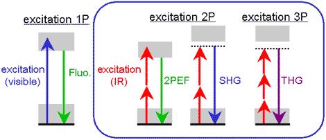
This latter goal is the focus of recent work by Beaurepaire and his colleagues. The methodology, developed during the thesis work of Willy Supatto and Delphine Débarre and used for this project, involves the following: femtosecond infrared pulses (one millionth of a billionth) focused within the embryo induce localized photoionization at the focal point via nonlinear interaction with the tissue. By controlling the deposited energy, it is possible to destroy regions ranging from hundreds of nanometers to hundreds of micrometers without disturbing neighboring cells. Additionally, less energetic pulses generate a third-harmonic signal upon encountering optical heterogeneities within the embryo. This multiphoton microscopy technique provides a micrometric map of embryonic structures without the need for external labeling. By combining these images with velocity analysis algorithms used in fluid mechanics, a complete description of morphogenetic movements during development can be achieved.
The obtained results (1) illustrate how ultra-short laser pulses enable:
- three-dimensional targeted ablations in living Drosophila embryos that modulate certain morphogenetic movements,
- a description of velocity fields and tissue deformations during development without fluorescent labeling (non-invasive method).
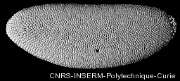
Figure: Drosophila embryo development visualized in 4D (3D + time) with two-photon (2PEF) microscopy using GFP in cell nuclei.
This research has deepened the investigation of twist’s mechanosensitive expression in stomodeal cells, essential for the proper formation of the future gastric system at the anterior pole in response to compression by the embryo's gastrulation convergence-extension movement. It quantitatively shows a strong correlation between this gene's biochemical expression pattern and the mechanical tissue deformation pattern deduced from a quantified analysis of in vivo morphogenetic movements.
This approach could be applied to other organisms; the ability to inactivate intra-embryonic structures via photo-ablation complements conventional genetic and biochemical approaches and could provide new insights into various developmental biology questions.